Published originally on Dental Economics
by Paul L. Child Jr., DMD, CDT, CEO CR Foundation
For more on this topic, go to www.dentaleconomics.com and search using the following key words: digital dentistry, dental technology, digital radiography, CAD/CAM, cone beam.
The connotations of being past the year 2010 bring about thoughts of futuristic concepts as suggested by movies, the Internet, and a vast array of media. Movies and books, set in a time period only a few decades forward, have portrayed a life filled with advanced medicine, travel, engineering, manufacturing, and even rapid and simple food production. Yet, when we reach that future date, we observe that technology doesn’t change as fast as our minds imagine. Does dentistry today — often termed “digital dentistry” — represent the high-tech, easy-to-implement solutions that were imagined and written about 30 years ago or even last year?
Clinicians with decades of experience or the student of dental history can look back at the advances in dentistry and state clearly that the dental profession has experienced an exciting amount of technological growth. Yet in comparison to medicine, biomedical engineering, automotive and aeronautics, rapid manufacturing, electronics, and others, dentistry appears to be more than a decade behind in adopting or integrating new technologies on a widespread basis. Although this statement may frustrate some early adopters and manufacturers of the new, available technologies in dentistry, a comparison of the technologies used in other advanced industries on a routine basis clearly demonstrates this chasm. If other industries have adopted newer and better technologies (including sharing them among one other), why does dentistry lag behind? Where does our profession stand with new technologies, and where might we be going?
The purpose of this article is to examine the concept of digital dentistry, its advantages and limitations, and make statements and observations on specific areas of digital dentistry based on research, direct personal experience, and communication with dental manufacturers and clinicians worldwide. It is intended to provide a practical view of digital dentistry, a stimulus for greater adoption of the areas that are proven, and faster integration of new technologies from which our profession can benefit.
 |
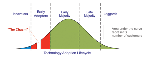
| Fig. 1 — Disruptive innovations and technology follow this curve. (Attributed to Craig Chelius; Technology Adoption Lifecycle as described in Geoffrey Moore’s book “Crossing the Chasm.”) |
What is digital dentistry?
Digital dentistry may be defined in a broad scope as any dental technology or device that incorporates digital or computer-controlled components in contrast to that of mechanical or electrical alone. This broad definition can range from the most commonly thought area of digital dentistry — CAD/CAM (computer aided design/CAD/CAM and intraoral imaging — both laboratory- and clinician-controlled
- Caries diagnosis
- Computer-aided implant dentistry — including design and fabrication of surgical guides
- Digital radiography — intraoral and extraoral, including cone beam computed tomography (CBCT)
- Electric and surgical/implant handpieces
- Lasers
- Occlusion and TMJ analysis and diagnosis
- Photography — extraoral and intraoral
- Practice and patient record management — including digital patient education
- Shade matching
There are many other areas of digital dentistry available, and many more are being researched. It is an exciting time to be in the dental profession as more technologies are being introduced that make dentistry easier, faster, better, and — most important — enjoyable.
Adoption and integration of technology
It took roughly two years for air rotor handpieces to gain widespread adoption and replace belt-driven handpieces, about five years for widespread adoption of PFM crowns, and about 25 years for implants. Why such a difference when all are now proven and widely used?
Some new technologies are “disruptive” in nature and can cause rapid change (see Fig. 1). The introduction of full zirconia crowns (BruxZir by Glidewell, and others) and other monolithic crowns (IPS e.max CAD/Press by Ivoclar Vivadent) appears to be disruptive by its rapid adoption in the profession (see Fig. 2).
The examination of other industries and past technological advances proves it generally takes up to 25 years for a new technology to be accepted and widely used (conversion from the early adopters to the early majority as in Fig. 1). If digital dentistry is now perceived as the future of dentistry, is it also behind by 25 years?
Dentistry, in comparison to the larger industries previously mentioned, is extremely small in terms of financial revenue, potential capital market growth, and outside, nondental investors. As such, some of the technological advances being developed in other industries are slow to be integrated in dentistry due to the relatively small global interest and financial input required to transfer the technology so that more efficient and improved dentistry results.
However, even though there are newer and better technologies being used in other industries, today dentistry is at the forefront of technology available within our industry, and more clinicians should become part of the early majority.
An integral part of understanding the future of dental technology involves observing and understanding new technologies in other industries and how that technology can be integrated into dentistry.
Advantages of digital dentistry
Each area of digital dentistry has advantages in comparison to the conventional device or technique. Yet, some of the advantages may be diminished by the increased cost or technique sensitivity.
As an example, although diode lasers have been available for more than a decade, early majority adoption did not occur until the recent decrease in prices of lasers and increased offerings and competition. This has resulted in an alternative to the lower cost electrosurgery devices.
 |
| Fig. 3 — Reconstructed 3-D image of the author (made with iCAT and Anatomage InVivo 5 software). 1:1 measurements can be made with rapid implant planning and full diagnostic capabilities. |
On the other hand, chairside intraoral imaging and fabrication of indirect restorations by the clinician have been available for more than 25 years (via CEREC by Sirona). However, even with new competition driving faster innovation (E4D by D4D Technologies), the price remains high, and adoption has not yet reached early majority (although it probably should have).
To be considered a clear advantage, the area of digital dentistry must include three things:
- Improved efficiency — both cost and time
- Improved accuracy in comparison to previous methods
- A high level of predictability of outcomes
Some areas of digital dentistry lack one or more of these characteristics and could easily be improved by adopting or integrating technology from other industries, or eliminating attempts to improve older, outdated technology and implementing newer, disruptive technology.
Limitations of digital dentistry
The major limitation of most areas of digital dentistry is cost. To adopt new technology often requires a higher capital investment, especially at the “innovator” or “early adopter” stage. Despite this, if the new technology meets the above criteria to be considered an advantage, then return on investment can be high if properly implemented.
One of the common pitfalls in adopting new dental technology is lack of desire on the part of the clinician and team to be adequately trained. Some clinicians will purchase a new technology, yet never read the owner’s manual or seek advanced training on how to operate the technology efficiently, often leading to high failure and abandonment. Misunderstanding the new technology tends to foster slower adoption rates. This scenario can be easily avoided by greater attendance of basic and advanced hands-on courses in these areas of technology — not just the state’s minimum to maintain the dental license.
Major areas of digital dentistry experience growth
I will comment briefly on the major areas of digital dentistry, their direction, and possible future developments. In determining your adoption and integration of some of the digital dentistry technologies into your practice, consider the following:
- What is your level of interest?
- Will the new technology add new excitement, drive, and happiness in your practice?
- Can you obtain a moderate to high return on investment?
- Are you willing to dedicate the time and effort to learn the new technology and continue advancing?
Practice and patient records management and patient education
Implementation of computers into each operatory and throughout the practice is the first and most frequent adoption of digital dentistry. In North America and most developed countries, this has reached the “early majority” stage as all of the criteria for being an advantage have been met.
Dentists who have not yet adopted this prerequisite for digital dentistry should do so now! Daily advances and improved software adapted from other industries allow this technology to be affordable, attain the fastest adoption rate, and offer a high return on investment. Current and highly effective systems include Eaglesoft (Patterson), Dentrix (Schein), PracticeWorks (Carestream Dental), and Web-based software such as Curve Dental.
Digital patient education is growing rapidly. The future in this area will reveal technologies and methods of communication already available in other industries, such as voice-activated and/or touch-screen computer and software instruction, live video and rapid recall of photos and educational components, 3-D video presentation with and without monitors or tablets, and off-site live consultation and education.
There are many effective options for digital patient education, including CAESY (Patterson), Guru (Schein), DDS GP for iPad (Kick Your Apps), Consult-PRO Chairside (Consult-PRO), etc. A recent CR Foundation (CLINICIANS REPORT) survey of more than 1,000 dentists revealed that 80% would consider purchasing an iPad or tablet for patient education. Clearly the time of drawing sketches on paper or using our hands to demonstrate teeth are a thing of the past.
Digital radiography
The next logical investment into digital dentistry (after full implementation of computers into your practice) is conversion to digital radiography. CLINICIANS REPORT and many other researchers have reported on the advantages of both intraoral and extraoral digital radiography.
The main advantages include lower radiation (when following the ALARA principle), significant time reduction, ease of storage and organization, and image enhancements for quick and improved viewing. Although the cost has not decreased significantly during the past five to eight years, the advantages far outweigh any limitations.
New and existing developments include wireless sensors (both CCD/CMOS and PSP), caries diagnosis (Logicon by Carestream Dental), Intelligent Positioning System for quick and easy digital alignment of the tube head to the sensor (Carestream Dental), and integration with tablets and voice activation.
Future improvements will use algorithms based on thousands of patient radiographs that accurately diagnose caries and make suggestions for the dentist. The potential for complete conversion to extraoral imaging only is a serious possibility for the future. There are currently many excellent intraoral digital radiography systems to choose from including Kodak, Dexis, Schick, Gendex, ScanX, etc.
Cone beam computed tomography
Cone beam CT is an exciting technology that has seen rapid growth due to decreased costs, many options to choose from, increased number of general dentists placing implants, decreased radiation compared to conventional CT scans, and rapid adoption by universities and specialists.
Although some states, provinces, and countries are struggling with how to regulate this fast-growing area of digital dentistry, its effectiveness and accuracy are unparalleled (see Fig. 3). Due to the moderate learning curve to understand the anatomy, software, and diagnostic capacity, dentists are encouraged to obtain further advanced education on this “disruptive” technology. When implemented properly, the return on investment for many clinicians is far superior to any other area of digital dentistry.
Cone beam CT is being quickly adopted by most specialties and becoming the proposed standard for many surgical procedures, including implant placement, third-molar removal, and endodontics. Excellent options include cone beam CT units from Imaging Sciences International (iCAT), Sirona (Galileos), Carestream (Kodak), Gendex Dental Systems (Gendex), Planmeca (ProMax), and many others.
Future advances and changes will see continued decrease in cost, improved software diagnostic capabilities to automatically take measurements and propose implant positions, algorithms that automatically look for assymetries and pathology to alert the radiologist for further examination, and rapid treatment planning for surgeries.
CAD/CAM and intraoral imaging
CAD/CAM for dental manufacturing and the dental laboratory profession is already in the early majority and will soon approach the late majority. The laboratory profession has discovered what the clinicians have been slower to recognize — CAD/CAM works. It is faster, more economical, predictable, consistent, and relatively accurate. Return on investment can be incredible if a team approach is adhered to.
CEREC has been available nearly 30 years, and recent advances with both CEREC and E4D clearly demonstrate that chairside CAD/CAM is uniquely positioned to lead our profession in digital dentistry. Merging of procedures, such as implant placement and immediate provisionalization, through strategic company alliances and shared technology allows dentists to do more in less time.
Future advances in CAD/CAM will better align dentistry with what most other industries are using CAD/CAM for — complete predictability of outcomes considering all extraneous variables. This would include automatic restoration design with no further modifications based on all patient factors, such as skeletal and arch classifications; wear, age, and tooth conditions; excursive movements; TMJ condition; exact input of condylar movements in relation to tooth positions; and design based on esthetics and desired look.
For these future advances to take place, manufacturers will need to further adopt and integrate technologies from other industries and create pathways for increasing investment by moving from “early adopters” to “early majority.”
For those who have sworn never to fabricate an indirect crown chairside or in their office, digital intraoral imaging/impressioning is growing rapidly and should draw every dentist’s attention. Scanning teeth and preparations is becoming increasingly easier and faster.
Currently, there are more than eight companies that offer intraoral imaging, with CEREC (Sirona), E4D (D4D Technologies), LAVA COS (3M), and iTero (Cadent/Align) being the most recognized and used. CR Foundation (CLINICIANS REPORT) has researched all of these scanning systems and proven all to be as accurate as the conventional methods (i.e., stone die systems). Most are more accurate, faster, and easier. It is not a question of, “Will CAD/CAM and intraoral imaging replace elastomeric impressions (i.e., VPS, polyether)?” but “When?”
Lasers
Diode lasers are one of the lowest costing areas of digital dentistry to adopt, as well as one of the easiest. Only in the past two years has the cost of diode lasers decreased to a level where “early majority” adoption is taking place.
Advantages of excellent hemostasis, universal use around all restorations, simplified surgical procedures, and an expanding use in a plethora of dental procedures make this area of digital dentistry highly desirable. The current trend is small, portable, cordless, low-cost diode lasers, such as the NV1 (Discus/Philips) and iLase (Biolase).
Other corded versions such as the Navigator (Ivoclar), EZlase 940 (Biolase), and Picasso (AMD) remain popular and effective. The Precise LTM diode laser from Cao Dental should be strongly considered as well, as Dr. Densen Cao is one of the originators and main innovators of diode lasers and LED curing lights.
Advances in lasers include the expanded use in almost every field of dentistry. Further research is needed to validate many of the claims, but many users of not only diode lasers but the other categories (CO2, Nd:YAG, erbium, etc.) have integrated lasers very effectively into their practices, and their observations seem to correlate with the claims.
Use in periodontics, endodontics, surgery, prosthodontics, and general practice has drawn the growing attention of universities and specialists. Future advances will see integration into dental operatory equipment, similar to LED curing lights and intraoral cameras, as well as other software hands-free control advances similar to that in other areas of digital dentistry.
Conclusion
Digital dentistry is more than just hype. When properly implemented and fully educated, return on investment can be excellent, increased joy in practicing dentistry can be experienced, and better care for your patients can be delivered.
The future of dentistry is now. Waiting another 10 years to adopt or integrate these new areas of dentistry will leave you decades behind. Decide which areas will best augment your practice, make informed decisions regarding your choice of product/technology, obtain education and training, and have fun!
Dr. Paul L. Child Jr. is a prosthodontist, certified dental technician, and CEO of CR Foundation (formerly CRA). Dr. Child lectures nationally and internationally on all areas of dentistry with an emphasis on new and emerging technologies. He maintains membership in many professional associations and academies, and is on the editorial board of several journals. Contact him at toni@pccdental.com.
This tutorial explains how to prepare a bridge or crown in Meshmixer ready for printing in any 3D printing device. It is generally a good idea to print the piece at a 30 to 45 degree tilt.
This explains how to close the mesh generated by a 3D scan of a model ready for 3D printing. It is important to close the mesh, otherwise it cannot be printed.
En este video tutorial vamos a dejar listo Meshmixer para que tengamos una referencia sobre el tamaño de bandeja de la DentalFab

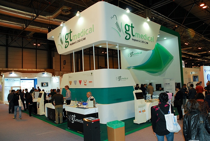
GT MEDICAL is a spanish company specialized in CAD/CAM applied to implantology that will be the exclusive distributors of the DentalFab in Spain and Portugal. GT Medical has more than 10 years of experience and they stand for the development of the Scan-Fit 1 and 2 scanner, both along with their own development CNC milling machine, will be part of a turn-key solution for the digital dental lab.
La impresión 3D está llamada a revolucionar la industria. Estudios de arquitectura y empresas de ingeniería ya se están beneficiando en gran medida de este adelanto. Sin embargo, aunque mucho más desconocidas, esta tecnología tiene numerosas y muy sorprendentes aplicaciones en el ámbito de la medicina.
Algunos de estos usos están todavía en una fase experimental, como la extrusión de células vivas (en lugar del habitual material plástico), que ha dado lugar a la bioimpresión de tejidos. Sin embargo otros se están generalizando: cada vez es más normal la impresión 3D de prótesis e implantes. He aquí un repaso por las ocho principales aplicaciones de la impresión 3D para la medicina.
Aúdifonos
El primer ámbito sanitario donde llegó este avance, aún antes incluso de que se hablase de impresión 3D fue el de las prótesis auditivas. Desde 1998 se vienen fabricando audífonos con máquinas precursoras de la impresora 3D. Con la generalización de este avance y las mejoras técnicas se ha llegado a un punto en el que el 98 por ciento de los aparatos se imprime con impresoras 3D y una sola máquina puede llegar a imprimir 30 aparatos en hora y media.
Prótesis dentales
Los dentistas fueron los pioneros en la generación de implantes a través de impresión 3D, una disciplina que es la que más está creciendo y en la que se obtienen algunos de los avances más llamativos. Este tipo de tecnología aplicada a la odontología ha aumentado la calidad de las piezas y acelera la producción.
Implantes óseos
Si hasta ahora se ha destacado lo que ya ha hecho por la medicina la impresión 3D, mucho más sorprendente es las posibilidades que abre de cara al futuro y que ya se están comenzando a llevar a cabo. Una de ellas es la posibilidad de crear implantes de hueso más compatibles y personalizados a un bajo coste. Recientemente, una niña de tres años con hidrocefalia salvó su vida en China al serle trasplantado un cráneo de titanio obtenido con esta técnica.
Trasplantes cardiacos
Hace un año, el Hospital de La Paz anunció que su área de Cardiología Pediátrica está trabajando junto a socios tecnológicos como Siemens para desarrollar mediante impresión 3D prótesis cardiacas personalizadas y adaptadas a cada paciente. En este convenio de colaboración científico-tecnológica participa el instituto de investigación del centro, el IdiPaz, junto con los profesionales del área y la compañía alemana.
Rehabilitación
El ingeniero español Juan Monzón ha unido impresión 3D con otra tecnología innovadora, los dispositivos wearables, para crear Exovite, un sistema de rehabilitación ósea y muscular. Este proyecto utiliza las impresoras 3D para crear férulas a medida del paciente, que inmovilizan el miembro fracturado e incorporan un sistema de electroestimulación que, conectado al móvil, permite seguir la rehabilitación desde casa, lo que reduce el tiempo de recuperación y los costes. El Hospital MAZ de Zaragoza será el primero en aplicar esta innovadora técnica en sus pacientes.
Simulacros para cirugía
Una de las aplicaciones más destacadas de esta tecnología la han encontrado los traumatólogos. Se trata de la creación de modelos de los huesos que van a operar. De esa manera, se evita la improvisación en los procedimientos quirúrgicos, se reducen los tiempos en quirófano y la recuperación de los pacientes es mucho más rápida. El proceso implica obtener una imagen completa de la lesión con un TAC y enviar los datos a un ordenador para que éste imprima el modelo. Cuando se habla de impresión 3D parece que se dan por supuesto inversiones elevadisimas, pero el Gregorio Marañón, por ejemplo, está haciendo esto valiéndose con dispositivos domésticos.
Impresión de tejidos
La llamada bioimpresión es un procedimiento enfocado al desarrollo de piel y tejidos impresos en 3D. En relación con la piel, la Universidad de Leiden (Holanda) está desarrollando un proceso, combinando esta tecnología y las llamadas células madre inducidas (iPS) para crear este tejido. Otra vía es la que está estudiando la Facultad de Medicina de la Universidad Wake Forest, donde están desarrollando una impresora 3D para fines militares, que puede imprimir piel directamente en pacientes quemados. La “tinta” está fabricada con diferentes tipos de células epiteliales. Se puede ver en la web de la universidad.
Impresión de órganos
De momento es la aplicación más alejada de las posibilidades actuales y que parece más ciencia ficción. Sin embargo, la impresión de células de hígado es algo que ya se está llevando a cabo e investigando para la experimentación con fármacos. La compañía estadounidense Organovo trabaja en intentar crear estos órganos, pero en miniatura, con un espesor de solo medio milímetro y 4 milímetros de anchura. Este experimento es importante para, en un futuro, crear estructuras de mayor tamaño, incluidas las redes de vasos necesarias para nutrirlas.


Recent Comments